Comparison of TGF-β and Type 1 Collagen Expression between Platelet Rich Fibrin Membrane and Conjunctival Autograft Treatment after Conjunctival Excision (An Experimental Animal Model Study)
Doi: 10.36351/pjo.v40i3.1791
DOI:
https://doi.org/10.36351/pjo.v40i3.1791Abstract
Purpose: To compare TGF- β and type 1 collagen expression between Platelet rich fibrin (PRF) membrane and conjunctival autograft treatment after conjunctival excision.
Study Design: Experimental animal study.
Place and Duration of study: Faculty of Veterinary Medicine, Airlangga University, Surabaya, Indonesia from October 24, 2022 to November, 19, 2022
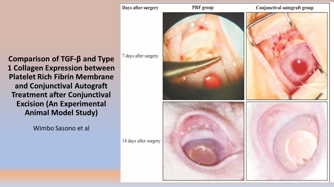
Methods: Twenty New Zealand white rabbits were randomly assigned to either the PRF group or the conjunctival autograft group. A 5x5 mm excision was made in the temporal quadrant of the right eye of each rabbit. In the first group, the conjunctival defect was closed using a PRF membrane, while in the second group, closure was done with a conjunctival autograft from the superior quadrant of the same eye. After 14 days, all rabbits were terminated and enucleated. An immunohistochemical study of conjunctival tissue was conducted to assess TGF-β and type 1 collagen expression, and results were statistically analyzed.
Results: An independent T-Test revealed that PRF membrane group exhibited higher TGF-β expression compared to the conjunctival autograft group (p = 0.000), with a mean TGF-β expression of 9.02 in the PRF membrane group and 5.76 in the conjunctival autograft group. Conversely, type 1 collagen expression was found to be higher in the conjunctival autograft group compared to the PRF membrane group (p = 0.032), with a mean type 1 collagen expression of 9.22 in the conjunctival autograft group and 6.92 in the PRF membrane group.
Conclusion: TGF-β expression was higher in the PRF group and type 1 collagen expression was higher in the conjunctival autograft group.

Downloads
Published
How to Cite
Issue
Section
License
Copyright (c) 2024 Ferry Setiawan

This work is licensed under a Creative Commons Attribution-NonCommercial 4.0 International License.


