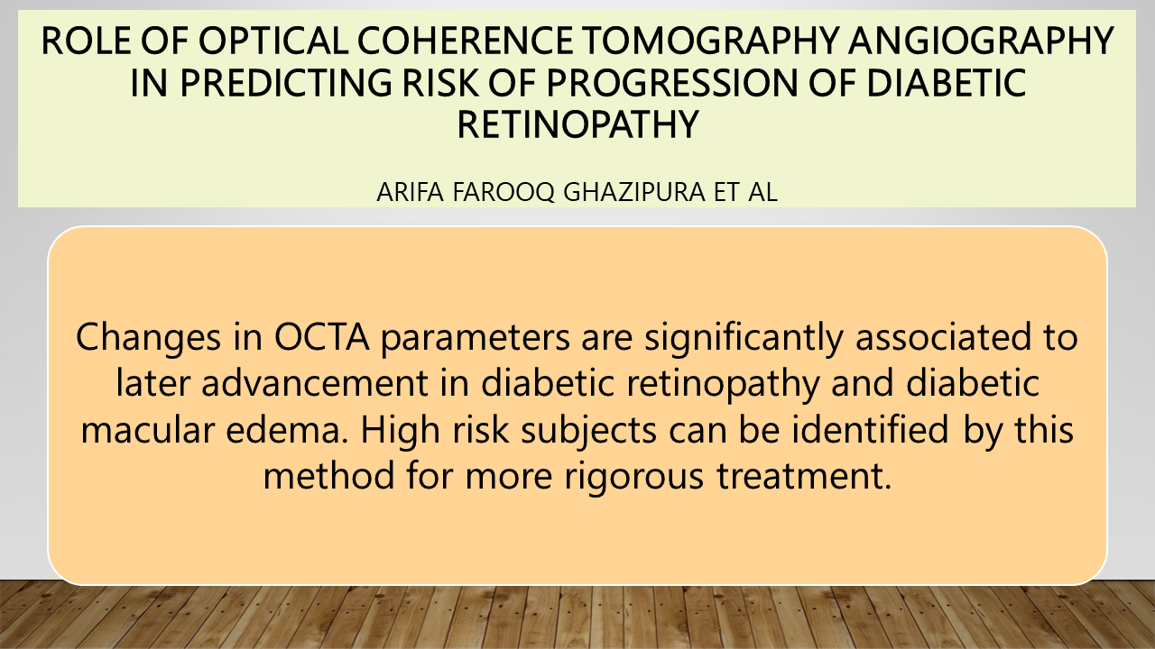
Role of Optical Coherence Tomography Angiography in Predicting Risk of Progression of Diabetic Retinopathy
Doi: 10.36351/pjo.v40i2.1718
DOI:
https://doi.org/10.36351/pjo.v40i2.1718Abstract
Purpose: To determine the role of Optical Coherence Tomography Angiography (OCTA)in predicting risk of progression of diabetic retinopathy.
Study Design: Descriptive observational study.
Place and Duration of Study: Jinnah Post graduate Medical Center, Karachi, from June 2022 to June 2023.
Methods: Patients with type 1 or 2 diabetes were included. Base line investigations were done including Central subfield thickness and OCTA at first visit. Second visit was conducted at 6th month and patients were followed for 24 months.OCT images of poor quality, motion artifact, inaccurate partition of tissue layers and blurry images were excluded.
Results: Among 97 enrolled patients, 88 cases finished the complete 24-months follow up. Out of 88 patients, 16 (18.18%) patients showed progression of diabetic retinopathy (DR), 9 (10.22%) patients developed DME, two showed both DR and DME .In univariate analysis, greater FAZ area, reduced FAZ circularity, Fractal dimension (FD) and vessel density (VD) of the deep capillary plexus (DCP) were significantly related to the advancement in DR. Among these, FAZ area, VD, and FD remained significant in the multivariate analysis. Whereas, only decreased VD and FD of superficial capillary plexus (SCP) were significantly related to advancement in DR and DME.
Conclusion: Changes in OCTA parameters is significantly associated with progression of diabetic ocular complications including diabetic retinopathy and diabetic macular edema. High risk subjects can be identified by this method for more rigorous treatment.

Downloads
Published
How to Cite
Issue
Section
License
Copyright (c) 2024 Dr Arifa Ghazipura, Dr. Najeebullah Achakzai, Dr. Gaintry, Prof. Dr Aziz ur Rehman, Prof. Dr Alyscia Marium Cheema

This work is licensed under a Creative Commons Attribution-NonCommercial 4.0 International License.


